A) Flexor carpi radialis
B) Brachialis
C) Pronator teres
D) Brachioradialis
E) Palmaris longus
G) A) and B)
Correct Answer

verified
Correct Answer
verified
Multiple Choice
 -This figure shows a posterior view of the right thigh. What muscle does number 2 indicate?
A) Semitendinosus
B) Semimembranosus
C) Biceps femoris
D) Adductor magnus
E) Gluteus maximus
G) C) and D)
Correct Answer

verified
Correct Answer
verified
Short Answer
Typically, the more movable attachment of axial muscles is the superior attachment. For muscles in the limbs, the more moveable attachment is the ________ attachment.
Correct Answer

verified
Correct Answer
verified
Multiple Choice
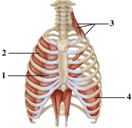
 -This figure shows the muscles of respiration. What muscles are indicated by number 3?
A) External intercostals
B) Internal intercostals
C) Scalenes
D) Diaphragm
E) Transversus thoracis
G) B) and E)
Correct Answer

verified
Correct Answer
verified
Multiple Choice
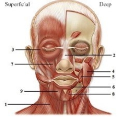
 -This figure shows the muscles of facial expression. What muscle does number 9 indicate?
A) Mentalis
B) Levator labii superioris
C) Risorius
D) Sternocleidomastoid
E) Depressor labii inferioris
G) All of the above
Correct Answer

verified
Correct Answer
verified
Multiple Choice
When contracted, this muscle causes expansion of the thoracic cavity and increases pressure in the abdominopelvic cavity. Its insertion is on a central tendon.
A) Internal intercostals
B) External intercostals
C) Transversus thoracis
D) Diaphragm
E) Serratus posterior inferior
G) A) and B)
Correct Answer

verified
Correct Answer
verified
Multiple Choice
All of the muscles listed, except one, are innervated by the deep fibular nerve. Select the exception.
A) Extensor digitorum longus
B) Gastrocnemius
C) Extensor hallucis longus
D) Tibialis anterior
E) Fibularis tertius
G) A) and B)
Correct Answer

verified
Correct Answer
verified
Multiple Choice
 -This figure shows superficial and deep arm muscles. What muscle does number 2 indicate?
A) Biceps brachii
B) Brachialis
C) Coracobrachialis
D) Brachioradialis
E) Triceps brachii
G) A) and C)
Correct Answer

verified
Correct Answer
verified
Multiple Choice
You just ran over a skunk on your way to class. The odor was overwhelming and in response you wrinkled your nose in disgust by contracting your
A) nasalis muscle.
B) procerus muscle.
C) depressor anguli oris.
D) frontal belly of the occipitofrontalis muscle.
E) occipital belly of the occipitofrontalis muscle.
G) B) and C)
Correct Answer

verified
Correct Answer
verified
Multiple Choice
Besides the supinator, which other muscle is a powerful supinator of the forearm?
A) Pronator teres
B) Pronator quadratus
C) Triceps brachii
D) Brachialis
E) Biceps brachii
G) A) and C)
Correct Answer

verified
Correct Answer
verified
Multiple Choice
For defecation to take place, the puborectalis must
A) contract.
B) relax.
D) undefined
Correct Answer

verified
Correct Answer
verified
Multiple Choice
The extensor digitorum muscle is found in the
A) deep layer of the posterior compartment of the forearm.
B) superficial layer of the posterior compartment of the forearm.
C) superficial layer of the anterior compartment of the forearm.
D) deep layer of the anterior compartment of the forearm.
F) B) and D)
Correct Answer

verified
Correct Answer
verified
Multiple Choice
These muscles elevate the ribs and have their origin on the inferior border of the superior rib and their insertion on the superior border of the inferior rib.
A) Internal intercostals
B) External intercostals
C) Transversus thoracis
D) Diaphragm
E) Serratus posterior inferior
G) B) and E)
Correct Answer

verified
Correct Answer
verified
Multiple Choice
Which muscle does not attach proximally to the ischial tuberosity?
A) Biceps femoris
B) Semimembranosus
C) Adductor longus
D) Semitendinosus
E) Quadratus femoris
G) D) and E)
Correct Answer

verified
Correct Answer
verified
Multiple Choice
The superficial layer of the urogenital triangle contains three muscles. Select the exception.
A) Puborectalis
B) Bulbospongiosus
C) Ischiocavernosus
D) Superficial transverse perineal
F) A) and C)
Correct Answer

verified
Correct Answer
verified
Multiple Choice
When a child raises her hand to show you she is five years old, she is using all of the following muscles except the
A) extensor digitorum.
B) flexor digitorum.
C) palmar interossei.
D) lumbricals.
E) dorsal interossei.
G) All of the above
Correct Answer

verified
Correct Answer
verified
Multiple Choice
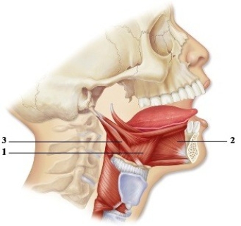
 -This figure shows the muscles that move the tongue. What muscle does number 1 indicate?
A) Palatoglossus
B) Styloglossus
C) Stylohyoid
D) Hyoglossus
E) Genioglossus
G) A) and D)
Correct Answer

verified
Correct Answer
verified
Short Answer
The rectus abdominis inserts on the ________ process, as well as on ribs 5-7.
Correct Answer

verified
Correct Answer
verified
Multiple Choice
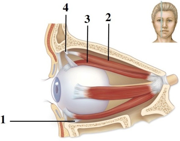
 -This figure shows a medial view of the right eye. What structure does number 4 indicate?
A) Trochlea
B) Common tendinous ring
C) Optic nerve
D) Optic canal
E) Superior rectus muscle
G) All of the above
Correct Answer

verified
Correct Answer
verified
Multiple Choice
If you had all of your fingers (including the thumb) spread out wide, which muscle or group would bring your thumb toward your first finger?
A) Adductor pollicis
B) Palmar interossei
C) Dorsal interossei
D) Lumbricals
E) Abductor pollicis longus
G) B) and E)
Correct Answer

verified
Correct Answer
verified
Showing 81 - 100 of 183
Related Exams