A) The calcaneus articulates with the tibia to form this joint.
B) Most common injuries to this joint occur because of a forceful inversion of the foot.
C) A capsule of hyaline cartilage surrounds the joint.
D) The lateral collateral ligament helps to stabilize this joint.
E) It is a pivot joint.
G) A) and D)
Correct Answer

verified
Correct Answer
verified
Multiple Choice
Tracie is experiencing pain on the bottom of her feet and her physician explains that it is due to ligament damage. Which of the following is most likely involved in Tracie's condition?
A) Plantar calcaneonavicular ligament
B) Transverse acetabular ligament
C) Interosseous membrane
D) Lateral collateral ligaments
F) A) and B)
Correct Answer

verified
Correct Answer
verified
Multiple Choice
Nick is climbing a rope at his local gym. He uses his hands and his feet to move up the rope. To "hold" on to the rope with his feet requires ________.
A) dorsiflexion
B) inversion
C) medial excursion
D) opposition
E) retraction
G) A) and B)
Correct Answer

verified
Correct Answer
verified
Multiple Choice
Arthritis is
A) a bacterial infection transmitted by ticks.
B) an inflammation of any joint.
C) a metabolic disorder caused by increased uric acid in blood.
D) a condition that may involve an autoimmune disease.
E) the most common type of arthritis.
G) A) and C)
Correct Answer

verified
Correct Answer
verified
Multiple Choice
With the elbow and wrist extended, painting a circle on a canvas requires ________ of the shoulder.
A) rotation
B) circumduction
C) extension
D) flexion
E) elevation
G) A) and B)
Correct Answer

verified
Correct Answer
verified
Multiple Choice
The joint capsule
A) is a double layer of tissue that encloses a joint.
B) is a thin lubricating film covering the surface of a joint.
C) provides a smooth surface where bones meet.
D) is a layer of tissue that is continuous with the periosteum.
E) lines the joint everywhere except over the articular cartilage.
G) D) and E)
Correct Answer

verified
Correct Answer
verified
Multiple Choice
A synchondrosis contains ________ cartilage.
A) synchronous
B) fibrous
C) elastic
D) reticular
E) hyaline
G) C) and E)
Correct Answer

verified
Correct Answer
verified
Multiple Choice
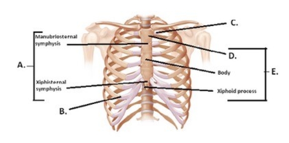
 -The figure illustrates the joints and bones of the rib cage. What does "B" represent?
A) Costochondral joint
B) Sternum
C) Manubrium
D) Sternal symphyses
E) Sternocostal synchrondrosis
G) A) and E)
Correct Answer

verified
Correct Answer
verified
Multiple Choice
The opposite of retraction is ________.
A) inversion
B) protraction
C) elevation
D) pronation
E) flexion
G) D) and E)
Correct Answer

verified
Correct Answer
verified
Multiple Choice
The ligament at the head of the femur is the ________.
A) ligamentum femoris
B) ligamentum teres
C) ligamentum acetabulum
D) ligamentum ilium
E) ligamentum primis
G) All of the above
Correct Answer

verified
Correct Answer
verified
Multiple Choice
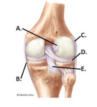
 -The figure illustrates a posterior view of the right knee joint. What does "D" represent?
A) Medial (tibial) collateral ligament (MCL)
B) Posterior cruciate ligament (PCL)
C) Anterior cruciate ligament (ACL)
D) Lateral (fibular) collateral ligament (LCL)
E) Lateral meniscus
G) B) and C)
Correct Answer

verified
Correct Answer
verified
Multiple Choice
Which of the following is mismatched?
A) Shoulder joint - coracohumeral ligament
B) Elbow joint - radial collateral ligaments
C) Hip joint - cruciate ligaments
D) Knee joint - patellar ligaments
E) Ankle - calcaneofibular ligament
G) None of the above
Correct Answer

verified
Correct Answer
verified
Multiple Choice
 -The figure illustrates a posterior view of the right knee joint. What does "A" represent?
A) Medial (tibial) collateral ligament (MCL)
B) Posterior cruciate ligament (PCL)
C) Anterior cruciate ligament (ACL)
D) Lateral (fibular) collateral ligament (LCL)
E) Lateral meniscus
G) B) and D)
Correct Answer

verified
Correct Answer
verified
Multiple Choice
An example of a saddle joint is the ________ joint.
A) shoulder
B) elbow
C) atlanto-occipital
D) carpometacarpal
E) atlantoaxial
G) A) and D)
Correct Answer

verified
Correct Answer
verified
Multiple Choice
Abnormal forced extension beyond normal range of motion is called ________.
A) circumduction
B) rotation
C) hyperextension
D) supination
E) pronation
G) A) and E)
Correct Answer

verified
Correct Answer
verified
Multiple Choice
The opposite of supination is ________.
A) inversion
B) protraction
C) elevation
D) pronation
E) flexion
G) A) and E)
Correct Answer

verified
Correct Answer
verified
Multiple Choice
Which of the following joints is most movable?
A) Suture
B) Syndesmosis
C) Symphysis
D) Synovial
E) Synchondrosis
G) A) and D)
Correct Answer

verified
Correct Answer
verified
Multiple Choice
The fibrous capsule
A) is a double layer of tissue that encloses a joint.
B) is a thin lubricating film covering the surface of a joint.
C) provides a smooth surface where bones meet.
D) is a layer of tissue that is continuous with the periosteum.
E) lines the joint everywhere except over the articular cartilage.
G) A) and E)
Correct Answer

verified
Correct Answer
verified
Multiple Choice
Which of the following movements is an example of extension?
A) Bending forward at the waist
B) Kneeling
C) Raising your arm laterally
D) Using your finger to point out an area on a map
E) Shrugging your shoulders
G) A) and E)
Correct Answer

verified
Correct Answer
verified
Multiple Choice
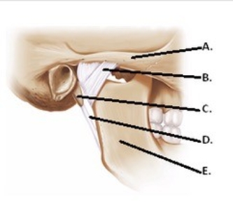
 -The figure illustrates structures in the right temporomandibular joint (lateral view) . What does "C" represent?
A) Lateral ligament
B) Mandible
C) Zygomatic arch
D) Styloid process
E) Stylomandibular ligament
G) B) and E)
Correct Answer

verified
Correct Answer
verified
Showing 101 - 120 of 149
Related Exams