Correct Answer

verified
Correct Answer
verified
Multiple Choice
The right border of the heart is supplied by the
A) circumflex artery.
B) posterior interventricular artery.
C) anterior interventricular artery.
D) right marginal artery.
E) great cardiac vein.
G) A) and E)
Correct Answer

verified
Correct Answer
verified
True/False
Because of the constant inflow of blood,the atria are thick-walled and located inferiorly in the heart.
B) False
Correct Answer

verified
Correct Answer
verified
Short Answer
The pulmonary arteries carry _______________ blood to the lungs.
Correct Answer

verified
Correct Answer
verified
Multiple Choice
During ventricular systole
A) only the AV valves open.
B) only the AV valves close.
C) only the semilunar valves close.
D) the semilunar valves close and the AV valves open.
E) the semilunar valves open and the AV valves close.
G) None of the above
Correct Answer

verified
Correct Answer
verified
Multiple Choice
Pectinate muscles are found on the
A) anterior wall of the right atrium.
B) posterior wall of the right ventricle.
C) anterior wall of the right ventricle.
D) anterior wall of the right and left atria.
E) posterior wall of the right and left ventricles.
G) B) and E)
Correct Answer

verified
Correct Answer
verified
Multiple Choice
Which analogy fits the human heart?
A) It is like a single pump.
B) It is like a double pump,each working side by side with the other.
C) It is like four pumps working in alternating cycles.
D) It is like a double pump,each working at its own rate determined by the needs of the body served.
E) It is like a single pump whose various chambers all work together at once.
G) A) and B)
Correct Answer

verified
Correct Answer
verified
Multiple Choice
Which describes the endocardium? A: Has single layer of epithelium B: Has layer of areolar connective tissue C: Epithelial cells are squamous D: Epithelial cells are cuboidal E: Has layer of adipose connective tissue F: Has patches of myocardium
A) a,b,c
B) a,b,d
C) a,d,e
D) a,b,c,e
E) a,e,f
G) D) and E)
Correct Answer

verified
Correct Answer
verified
Multiple Choice
The internal wall surface of each ventricle displays large,smooth,irregular muscular ridges called
A) conus arteriosus.
B) atrioventricular opening.
C) trabeculae carneae.
D) chordae tendineae
E) pectinate muscles
G) C) and D)
Correct Answer

verified
Correct Answer
verified
Short Answer
The property that allows the heart to initiate each heartbeat itself is called __________________.
Correct Answer

verified
Correct Answer
verified
Multiple Choice
The heart valves
A) stabilize and hold the arteries leaving the heart.
B) permit the passage of blood in one direction.
C) separate the right and left sides of the heart.
D) are only used in the fetal heart.
E) direct the conduction impulse through the heart muscle.
G) A) and D)
Correct Answer

verified
Correct Answer
verified
Short Answer
The inferior,conical end of the heart is called the _______________.
Correct Answer

verified
Correct Answer
verified
Multiple Choice
Cardiac muscle fibers
A) contract as a single unit.
B) are only loosely connected by the intercalated discs.
C) have a low oxygen need.
D) utilize hemoglobin as an energy source.
F) B) and C)
Correct Answer

verified
Correct Answer
verified
Multiple Choice
Which values are reasonable for a healthy,80 kilogram resting adult?
A) 5.25 liters of blood pumped per ventricle per minute;108,000 beats per day
B) 4.25 liters of blood pumped per ventricle per minute;60,000 beats per day
C) 6.75 liters of blood pumped per ventricle per minute;20,000 beats per day
D) 7.25 liters of blood pumped per ventricle per minute;76,000 beats per day
E) 5.75 liters of blood pumped per ventricle per minute;144,000 beats per day
G) B) and C)
Correct Answer

verified
Correct Answer
verified
Multiple Choice
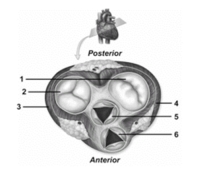
 -In this figure showing an oblique section of the heart,number 3 depicts the
A) wall of the left ventricle.
B) wall of the right atrium.
C) wall of the left atrium.
D) wall of the right ventricle.
E) left atrioventricular valve.
G) B) and D)
Correct Answer

verified
Correct Answer
verified
Multiple Choice
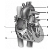
 -In this figure showing an anterior view of the heart,number 7 depicts the
A) aortic semilunar valve.
B) right atrium.
C) left ventricle.
D) right atrioventricular valve.
E) pulmonary semilunar valve.
G) A) and C)
Correct Answer

verified
Correct Answer
verified
Multiple Choice
It is the _____________ that permits the compression necessary to pump large volumes of blood out of the ventricles.
A) negative pressure inside the ventricles
B) absence of oxygenated blood in the atria
C) arrangement of cardiac muscle in the heart wall
D) presence of skeletal muscle tissue in the heart skeleton
E) presence of papillary muscles in the ventricles
G) None of the above
Correct Answer

verified
Correct Answer
verified
Short Answer
The relaxation phase of a heart chamber is termed ________________.
Correct Answer

verified
Correct Answer
verified
Short Answer
The heartbeat is initiated by the cardiac muscle fibers of the _______________ node.
Correct Answer

verified
Correct Answer
verified
Multiple Choice
The serous fluid within the pericardial cavity works to
A) lubricate the membranes of the serous pericardium.
B) slow the heart rate.
C) equalize the pressure in the great vessels.
D) eliminate blood pressure spikes.
F) A) and B)
Correct Answer

verified
Correct Answer
verified
Showing 81 - 100 of 101
Related Exams